
Figure 6.
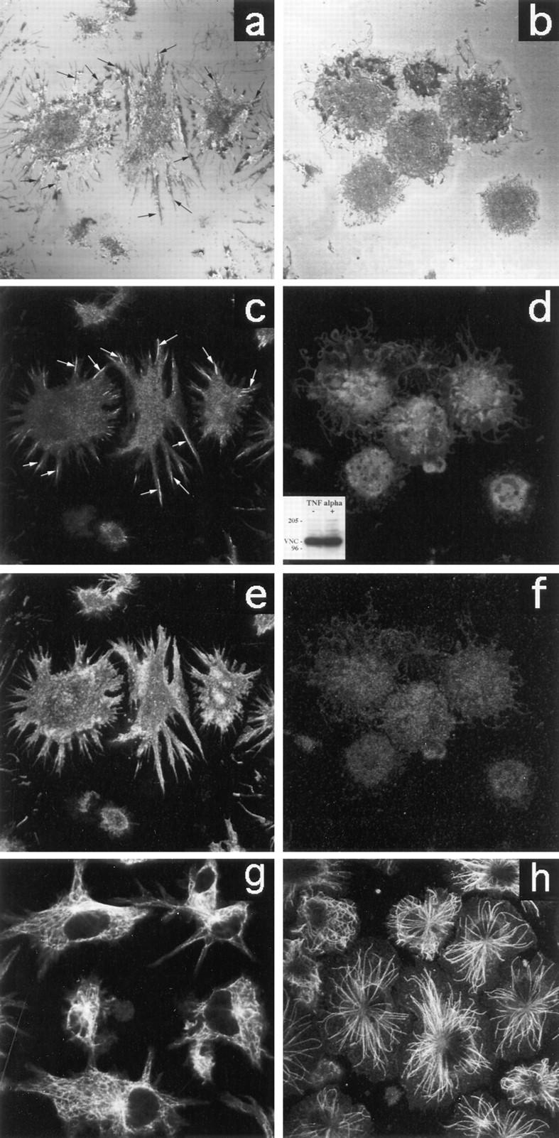
Cytoskeleton modifications in D1 cells after TNFα treatment. Immature (a) and mature (b) D1 cells morphology was analyzed by confocal microscopy using reflection interference contrast which gives the best visualization of the cell interface zone when attached to a glass support (37). Confocal laser scanning microscopy of D1 cells stained with anti-vinculin (c and d), phalloidin (e and f), and anti-tubulin (g and h) was also performed. Immature D1 cells (a, c, e, and g) appear to be adherent (a) and characterized by: vinculin containing adhesive structures (a and c, arrows), subcortical actin aggregates (e), and highly organized tubulin (g). D1 treatment with TNFα (b, d, f, and h) clearly induces morphological modification (b). Mature D1 cells lose adherence (b), vinculin (d), and subcortical actin organization ( f ). No differences in the amount of vinculin protein are detectable by Western blot analysis (d, inset). Microtubules are only partially affected by TNFα treatment (h).