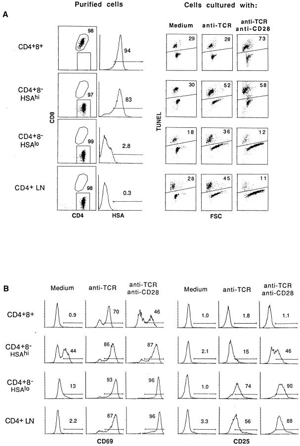
Figure 2.
Apoptosis of thymocyte subsets and LN T cells induced by stimulation with anti-TCR mAb or anti-TCR plus anti-CD28 mAbs in vitro. Purified populations of CD4+8+, HSAhi CD4+8−, and HSAlo CD4+8− thymocytes and CD4+8− LN cells were cultured for 20 h in vitro on plates coated with anti-TCR mAb alone (H57, 10 μg/ml) or with a mixture of anti-TCR (10 μg/ml) and anti-CD28 (20 μg/ml) mAbs. (A) The purity of the subsets before culture (left) and the extent of apoptosis determined by TUNEL staining after culture (right) are shown; for TUNEL staining, the forward scatter (FSC) profile shows the relative size of the cells. (B) Shows surface expression of CD69 and CD25 (IL-2R) on viable (TUNEL −) cells after culture. To exclude the possibility that CD4 ligation during the panning procedure affected the results (50), parallel experiments were performed with cells that were purified by methods that did not involve CD4 ligation, e.g., by using unmanipulated thymocytes from β2m −/− mice as a source of purified CD4+8+ cells and CD8− IL-2R− normal thymocytes as an enriched source of CD4+8− cells. The results obtained with these cells were essentially identical to the data shown in Figs. 3 and 4.

