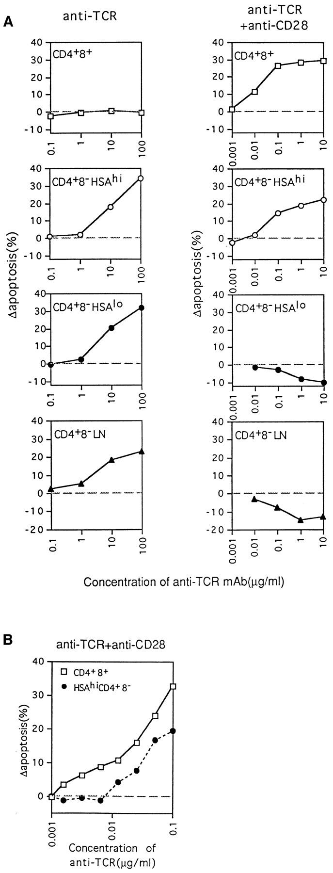
Figure 3.

Concentration of anti-TCR mAb required to induce apoptosis of thymocyte subpopulations as measured by TUNEL staining at 20 h. (A) Purified thymocyte subsets and LN CD4+8− cells were cultured on plates coated with various concentrations of anti-TCR mAb, i.e., 100 μg/ml to 0.1 μg/ml for stimulation with anti-TCR mAb alone, and 10 μg/ml to 1 ng/ml for stimulation with anti-TCR mAb plus a fixed amount of 20 μg/ml of anti-CD28 mAb. (B) Comparison of the sensitivity of CD4+8+ and HSAhi CD4+8− thymocytes to apoptosis induced by stimulation with various concentrations of anti-TCR mAb and a fixed amount (20 μg/ml) of anti-CD28 mAb. Background levels of apoptosis found with cells cultured in medium alone have been subtracted, and the data are shown as change in apoptosis. The data represent the mean from three (A) or two (B) separate experiments.
