Figure 7.
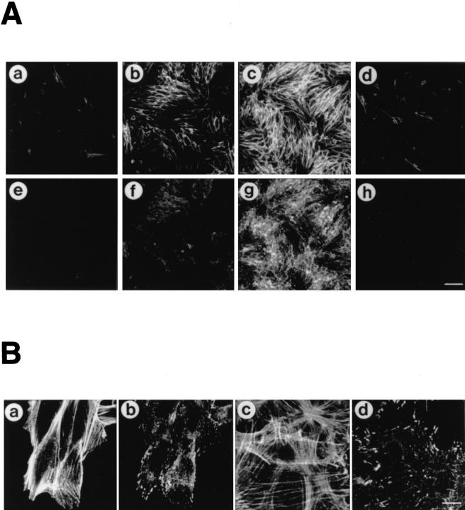
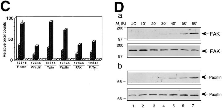
Rearrangements of F-actin and tyrosine phosphorylated proteins in CHO cells upon incubation in MEM containing secreted Ipa invasins. (A) Development of actin stress fibers and tyrosine phosphorylated proteins in CHO cells in response to addition of secreted Ipa invasins. Serum-starved CHO cells were incubated in MEM containing secreted Ipa invasins from S. flexneri YSH6000T for 0 min (a and e), 30 min (b and f), 60 min (c and g), or in MEM without secreted Ipa invasins for 60 min (d and h) at 37°C. Actin filaments were visualized with rhodaminephalloidin (a–d), and in the same cells phosphorylated protein tyrosine was localized by FITC-conjugated anti-phosphotyrosine (e–h). Bar, (h) 100 μm. (B) High magnification of A. Serum-starved CHO cells (a and b) and Swiss 3T3 cells (c and d) were incubated in MEM containing secreted Ipa invasins for 60 min at 37°C. Actin filaments were visualized with rhodaminephalloidin (a and c), and in the same cells phosphorylated protein tyrosine was localized by FITCconjugated anti-phosphotyrosine (b and d). Bar, (d) 10 μm. (C) Quantification of development of F-actin and assembly of focal adhesion proteins. Serum-starved CHO cells were incubated with secreted Ipa invasins for 0 min (lane 1), 30 min (lane 2), 60 min (lane 3), without Ipa invasins (lane 4) or were pretreated with C3 (1.25 μg/ ml) for 24 h (lane 5). Quantification of the pixel count was calculated using LaserSharp version 2.0 (Bio-Rad Labs.). Figures represent relative numbers compared with 0 min (lane 1). The data shown are the means of triplicate experiments. (D) Tyrosine phosphorylation of FAK and paxillin in serumstarved CHO cells upon incubation with MEM containing secreted Ipa invasins. Samples are from untreated cells (UC), and from cells incubated with Ipa invasins for up to 60 min. Cell lysate proteins were immunoprecipitated with the antiFAK mAb 2A7 (a) or anti-paxillin mAb 347 (b), separated by 10% SDSPAGE, and transferred to nitrocellulose, before probing with antiphosphotyrosine mAb PT-66 (a and b, top), anti-FAK mAb 2A7 (a, bottom), or anti-paxillin mAb 347 (b, bottom).


