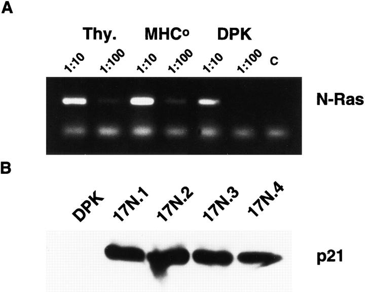
Figure 3.
Expression of a dominant negative mutant of p21ras blocks DPK cell differentiation. (A) RT-PCR assay of N-ras expression in DPK cells or thymocytes derived from wild-type or MHC-deficient mice. (B) Cell lysates from DPK cells and four independent lines that express Ha-ras N17 were analyzed by Western blot and probed with anti-ras antibody. Endogenous p21ras is not visible in this exposure. (C) DPK or 17N4 cells were cultured with DCEK-ICAM fibroblast antigen presenting cells in the presence (bold lines) or absence (thin lines) of 2 μM pigeon cytochrome c peptide. After 3 d of culture, cells were collected, stained with mAb to CD69 and analyzed by flow cytometry. (D) DPK or 17N4 cells were cultured as in (C) except that cells were harvested on day 1 or 3 as indicated and stained with anti-CD4 and anti-CD8 mAbs. Shown are the percentages of CD4+8lo/- DPK cells in the designated regions.


