
Figure 6.
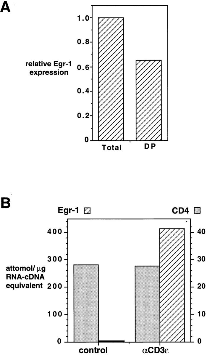
Expression of Egr-1 mRNA in double positive thymocytes. (A) Total thymocytes and CD4+8+ thymocytes (isolated by cell sorting, 95% DP) derived from the same animal, were assayed for expression of Egr and CD4 mRNA by competitive RT-PCR. Shown is the relative level of Egr-1 cDNA in the sample, normalized to the level of CD4 cDNA. (B) Total thymocytes derived from an MHC-deficient mouse were cultured with hamster immunoglobulin-coated or anti-CD3ε mAbcoated beads for 90 min before determination of Egr-1 and CD4 gene expression as in Fig. 1 C.