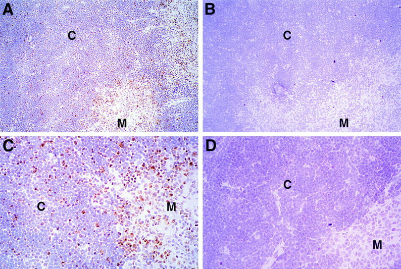
Figure 8.

Expression of Egr-1 protein in the thymus. Thin sections of normal thymus (A–C) or MHC knockout thymus (D) were fixed in formaldehyde and stained with a specific rabbit anti-Egr-1 peptide antiserum (A, C, D) or the same antibody preincubated with specific peptide (B). Regions of cortex (C) and medulla (M) are indicated. Sections were counterstained with hematoxylin and photographed at ×20 (A, B) or ×40 (C, D). C shows a magnification of the same section photographed in A.
