
Figure 2.
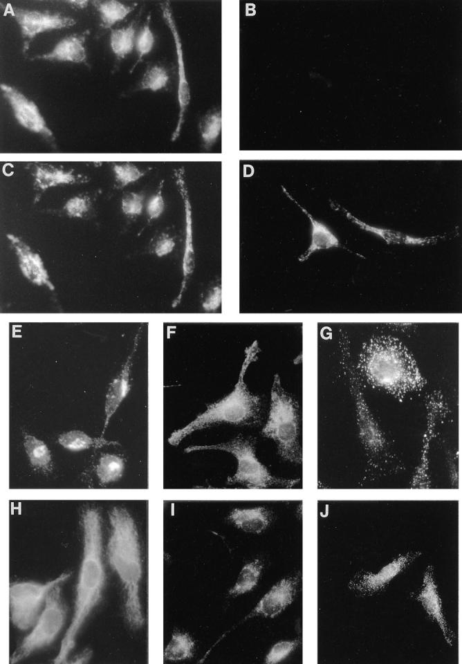
Colocalization of the Nramp1 and Lamp1 proteins in macrophages. Peritoneal macrophages were isolated from normal 129/sv (A, C, and E–J) and 129/sv Nramp1− /− mutants (B and D) and processed for double indirect immunofluorescence with the rabbit anti-Nramp1 antiserum GST-35C (A and B; 1:50 dilution) and a rat anti-Lamp1 monoclonal antibody (C and D; 1:10 dilution). Both cell populations were processed identically and equal exposure times were used for photography. Macrophages from 129/sv mice were also reacted with antibodies directed against MG160, a medial Golgi marker (E; 1:500 dilution) Rab5, an early endosomal marker (F; 1:200 dilution), cathepsins B and D, lysosomal proteases (G and J, respectively; 1:200 dilution), calnexin, a protein expressed in the endoplasmic reticulum and nuclear envelope (H; 1:100 dilution), and Rab7, a late endosomal marker (I; 1: 200 dilution).