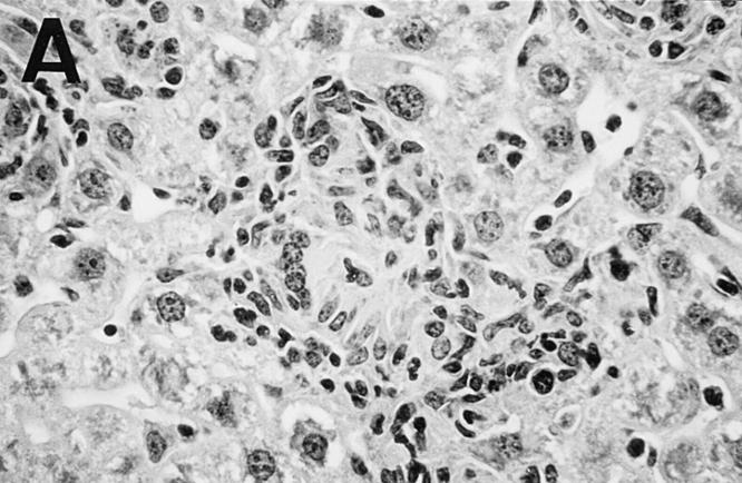
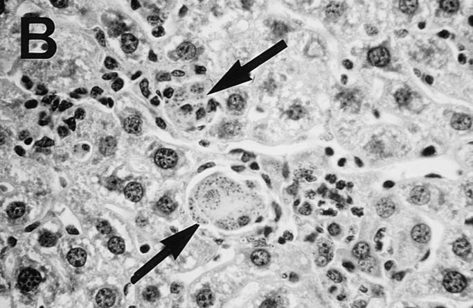
Figure 5.
Photomicrograph of liver sections from untreated control (A) and HKLMP-primed BALB/c mice (B) 4 wk after L. donovani infection. (A) shows a large, well-developed mature granuloma at an infected focus in normal mice. The granuloma contains few amastigotes. In B, HKLMP-primed mice show fused, heavily parasitized Kupffer cells (arrows), but little or no surrounding mononuclear cell infiltrate. ×200.


