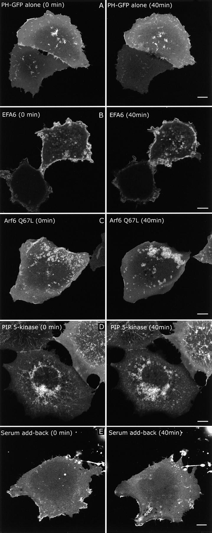
Figure 7.

Live cell dynamics of PIP2-labeled membranes after Arf6 activation. Cos cells were transfected with PH-GFP alone (A), EFA6 (B), or Arf6 Q67L (C) or PIP 5-kinase (D) and then imaged 18 h after transfection for ∼40 min. For serum add-back (E), PH-GFP–expressing cells were serum starved overnight. Serum (20%) was added immediately before imaging. Images of the subject cells taken at 0 and 40 min are shown. See also QuickTime videos 1–5 of each condition available at http://www.jcb.org/content/vol154/issue5. Each video shows the time points between the still images, are played at equivalent frame rates, and were recorded over the same approximate length of time. Bars, 10 μm.
