
Figure 3.
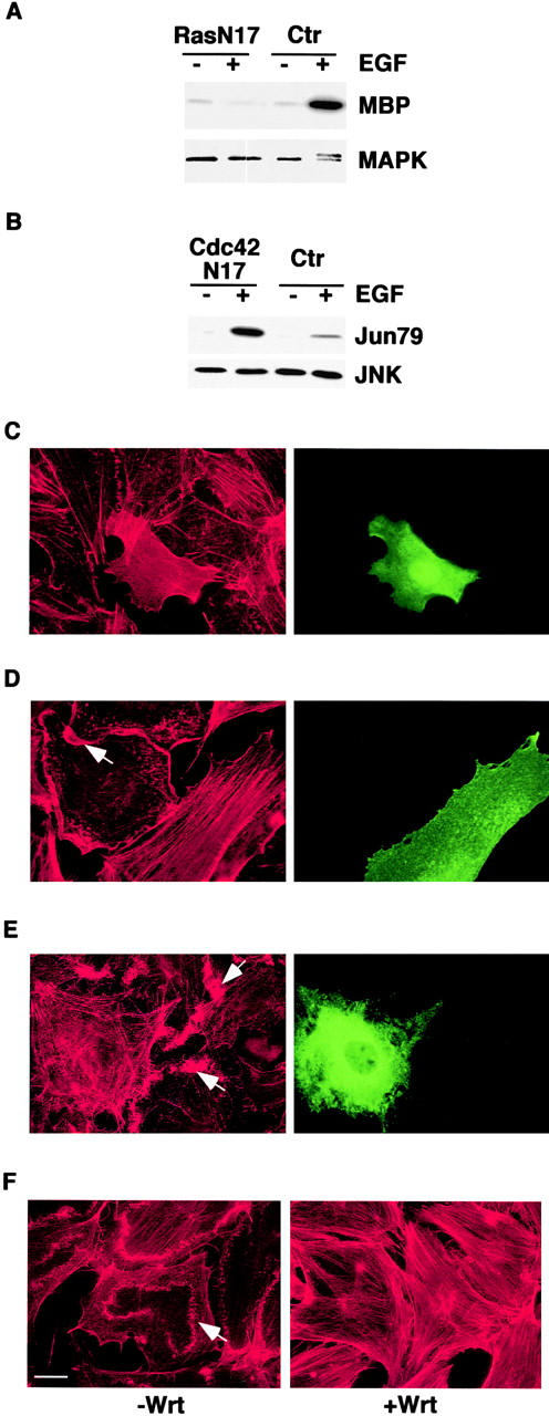
Expression and biological activity of dominant negative mutants of Ras, Cdc42, Rho, Rac, PI-3K, and of the PI-3K inhibitor, wortmannin. (A) Mouse embryo fibroblasts were cotransfected with an expression vector encoding RasN17 (Ras N17) or an empty vector as a control (Ctr), together with pCDNA-HA-MAPK. Cells were serum starved for 24 h and treated with 100 ng/ml of EGF (+) or mock treated (−) for 10 min. MAPK kinase activity was determined in immunocomplex kinase assays using myelin basic protein (MBP) as a substrate. An aliquot of the immunoprecipitates was also immunoblotted with anti-MAPK antibodies (MAPK). The doublet MAPK band, detected in the EGF-treated control sample, represents the active phosphorylated form. (B) Mouse embryo fibroblasts were cotransfected with expression vectors encoding a dominant negative HA-tagged Cdc42 (Cdc42N17) or an empty vector as a control (Ctr) together with pCDNA-HA-JNK, serum starved for 24 h, and treated with 100 ng/ml of EGF (+) or mock treated for 10 min (−). JNK kinase activity was determined in immunocomplex kinase assays, using the COOH-terminal region of c-JUN (Jun79) as a substrate. An aliquot of the immunoprecipitates was also immunoblotted with anti-JNK antibodies (JNK). The expression of RasN17 in A and HA-Cdc42N17 in B was determined by immunoblot analysis with anti-Ras and anti-HA antibodies, respectively (not shown). (C–E). Nuclei of quiescent mouse embryo fibroblasts were microinjected with expression vectors encoding RhoN19 (C), RacN17 (D), or p85ΔiSH2 (E). After 3 h, cells were stimulated with either 10% serum for 60 min (C) or PDGF (10 ng/ml) for 10 min (D and E) and fixed and stained with rhodamine-conjugated phalloidin (red) to detect F-actin, anti-Rho (C, green), anti-Rac (D, green) or anti-p85 (E, green) antibodies. (F) Quiescent mouse embryo fibroblasts were treated with wortmannin (100 nM) (+Wrt) or vehicle as a control (−Wrt) for 1 h before adding PDGF for 10 min. Cells were then fixed and stained with phalloidin (red). Arrows point to ruffles. Bar, 10 μm.