Figure 2.
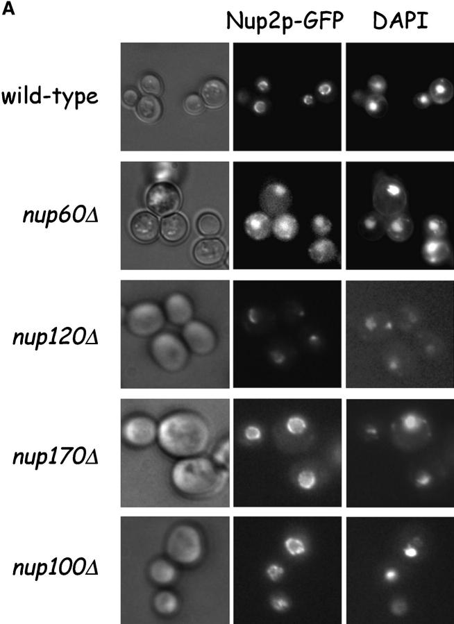
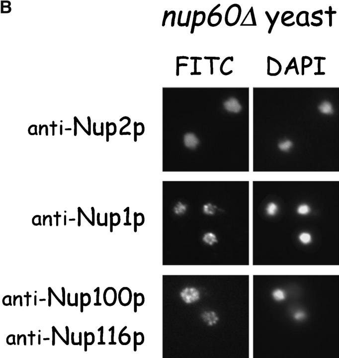
Nup2p is tethered to the NPC via Nup60p. (A) Direct visualization of Nup2p–GFP fusions in yeast. Various yeast strains that express NUP2-GFP from the NUP2 locus were grown in rich media at 30°C and were observed live under a fluorescence microscope. DAPI stain was used to visualize DNA in nuclei and pictures were taken using nuclei as the focal point. Note that in wild-type, nup170Δ, and nup100Δ yeast, Nup2p–GFP fusions accumulate in a punctate pattern at the nuclear periphery, but in nup60Δ yeast Nup2p–GFP is mislocalized to the nucleoplasm and cytoplasm. (B) Indirect immunofluorescence visualization of Nup2p, Nup1p, and Nup100p/Nup116p in nup60Δ yeast. nup60Δ yeast grown to early log phase in rich media at 30°C were fixed in 3.7% formaldehyde for 10 min and processed for immunofluorescence microscopy using affinity-purified anti-Nup antibodies and FITC-labeled secondary antibodies (left). DAPI was used to visualize nuclei (right). Note the mislocalization of Nup2p to the nucleoplasm in nup60Δ yeast in contrast to the normal punctate staining of Nup1p and Nup100p/Nup116p at the nuclear envelope.


