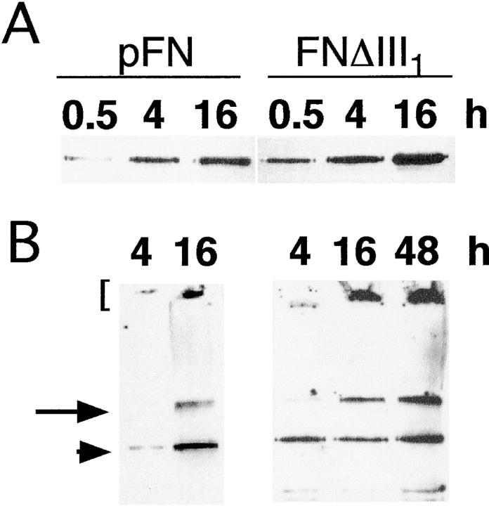
Figure 3.
DOC-soluble and -insoluble FNΔIII1. CHOα5 cells were incubated with 50 μg/ml pFN or FNΔIII1 for the indicated times and lysed in buffered DOC. DOC-soluble (A) and -insoluble (B) fractions were separated in 5% polyacrylamide-SDS gels without reduction and transferred to nitrocellulose. FN was detected on immunoblots with monoclonal anti-FN antibody IC3 and chemiluminescence reagents. Dimeric pFN and FNΔIII1 are present (arrowhead) as well as high molecular mass multimers at the top of the stacking (bracket) and at the interface of the stacking and separating gels (arrow).

