
Figure 9.
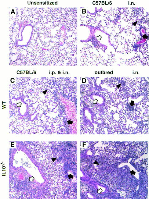
Lung lesions induced by sensitization with A. fumigatus Ag in outbred and C57BL/6 mice. Sections were prepared from formaldehyde-fixed lungs and stained with hematoxylin and eosin. Representative sections of a lung from each group of mice are shown (magnification, × 80). Lungs were removed 4 d after the second i.n. sensitization of outbred IL-10−/− (n = 9) and WT (n = 8) mice. Lungs were removed 4–7 d after the final i.n. challenge of C57BL/6 mice that had been primed i.n. or i.p. (n = 6–9). Open arrows point to peribronchial inflammation, closed arrows point to perivascular inflammation, and arrowheads point to alveolar lesions. (A) As lung sections from unsensitized WT and IL-10−/− mice were similar regardless of the mouse strain, one representative section is shown. (B) As lesions did not differ between WT or IL-10−/− C57BL/6 mice sensitized i.n., only one representative section is shown. (C and E) Lungs from C57BL/6 mice primed i.p. and challenged i.n. (C) WT, (E) IL-10−/−. (D and F) Lungs from outbred mice sensitized i.n. (D) WT, (F) IL-10−/−.