
Figure 3.
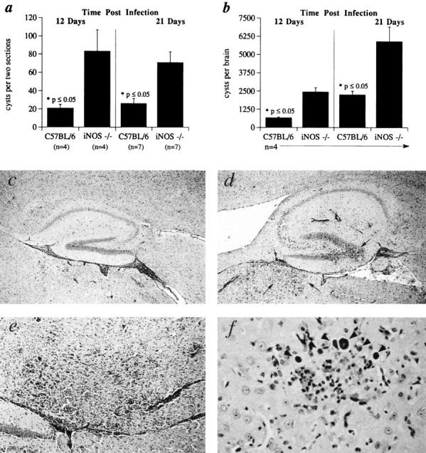
Brains of iNOS ko mice show increased cyst burdens as well as necrotizing encephalitis. C57BL/6 and iNOS ko mice were infected i.p. with 20 cysts each and brains harvested 12 and 21 d later for cyst enumeration in histological sections (A) or tissue homogenates (B). The mean cyst count and SE are shown for each group consisting of the number of animals indicated. (C–F) Photomicrographs of periodic acid–Schiff's stained, sagittal sections of brains from iNOS (D–F) and C57BL/6 (C) mice at 21 d after infection. Hippocampal regions of the brain in C57BL/6 (C) and iNOS ko (D) mice. Large areas of inflammation and necrosis seen in the iNOS brain (D) but not in C57BL/6 brain (C) are indicated by arrows. E shows a higher magnification of the same necrotizing lesion in D. (F) Brain section from iNOS ko mouse with numerous cysts (arrowheads) arranged in a satellite-like array, a feature frequently observed in these animals. Original magnification: (C and D) ×50; (E) ×200; (F) ×400.