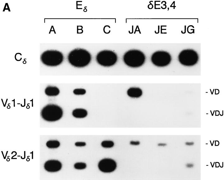
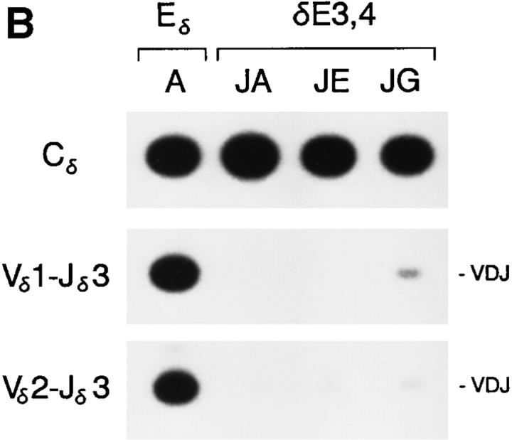
Figure 5.
PCR analysis of Eδ and δE3,4 minilocus rearrangement. Genomic DNA from thymi of wild-type Eδ mice from lines A, B, and C, and from δE3,4 mice from lines JA, JE, and JG were amplified by PCR using primers 5 and 6 (top), primers 1 and 3 (middle) and primers 2 and 3 (bottom). Southern blots were developed using radiolabeled Cδ, Vδ1, or Vδ2 cDNA probes. The mice analyzed were A-765 (4 wk old), B-31 (8 wk old), C-114 (4 wk old), JA-81 (4 wk old), JE101 (4 wk old), and JG-42 (4 wk old). (B) Genomic DNA from the thymi of mice from lines A, JA, JE, and JG were amplified by PCR using primers 5 and 6 (top), primers 1 and 4 (middle) and primers 2 and 4 (bottom). The mice analyzed were A-866, JA-81, JE-101, and JG-42 (all 4 wk old).


