
Figure 3.
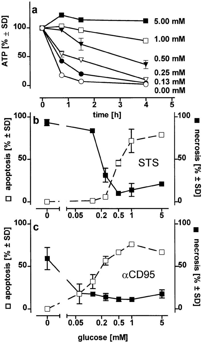

Changes in the mode of cell death after clamping the intracellular ATP concentration at defined levels or at different times. (a) Jurkat cells in glucose-free medium containing pyruvate (2 mM) were exposed to 2.5 μM oligomycin plus the indicated concentrations of glucose. Intracellular ATP concentrations were determined at the times indicated; (b and c) Jurkat cells were preincubated for 45 min in medium containing 2.5 μM oligomycin plus the concentration of glucose indicated. STS (b) or αCD95 (c) were then added and the mode of cell death was determined after further 3.5 h; (d ) intracellular ATP levels were manipulated during incubation of Jurkat cells with either STS or αCD95. Left, cells were incubated in pyruvate-supplemented medium without glucose and challenged (at t = 0) with 100 ng/ml αCD95 or 1.2 μM STS. At the times indicated oligomycin was added to deplete ATP, and the mode of cell death was determined 4 h after the challenge. Right, cells were first depleted of ATP by preincubation with oligomycin (at t = −1 h). At t = 0, STS or αCD95 were added. At the times indicated, intracellular ATP was replenished by adding 10 mM glucose to the incubation medium. The dashed bold line indicates the cellular ATP concentration (percentage of untreated control cultures in standard pyruvate medium), which was reached 15 min after each glucose supplementation. Data are means ± SD of triplicate determinations.