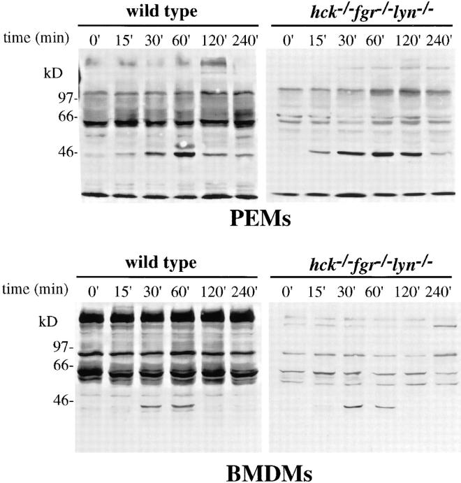
Figure 6.
Protein-tyrosine phosphorylation after LPS/IFN-γ stimulation. PEMs and BMDMs from wild-type and hck −/− fgr −/− lyn −/− mice were stimulated with LPS (10 ng/ml) and IFN-γ (20 ng/ml) for the different time points indicated. Total cell lysates were prepared and separated on 10% SDS-PAGE and transferred to nitrocellulose as described in Materials and Methods. Filters were blotted with antiphosphotyrosine with 4G10, followed by peroxidase-conjugated secondary antibody and chemiluminescent detection.

