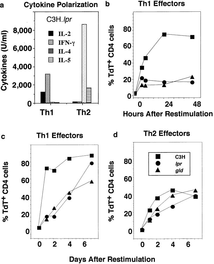
Figure 6.
Role of Fas and FasL in effector AICD. Th1 and Th2 effector cells were generated in vitro from naive CD4+ cells obtained from C3H (▪), C3H.MRL-Faslpr (•), and C3H/HeJ-FasLgld (▴) mice by culture with Con A (2 μg/ml) and DCEK-ICAM APC in the presence of IL-2 plus either IL-12 and anti–IL-4 (11B11) or IL-4 and anti–IFN-γ, respectively. After 5 d, well-polarized Th1 or Th2 effectors (5 × 10⁵/ml) were recovered and stimulated with Con A (2 μg/ml) and APC (1.7 × 10⁵/ml) again for hours or days as indicated. At the various times after restimulation, cultures were harvested. Viability was determined and samples were stained with TdT as described above. (a) Cytokine polarization. In parallel cultures, the effector populations were restimulated as above and supernatants were harvested 18–24 h after restimulation for determination of cytokine titers. The cytokine pattern of Th1 and Th2 effectors from wild-type C3H and C3H.lpr mice were very similar to those from gld mice (not shown). IL-2 was tested by bioassay, and IFN-γ, IL-4, and IL-5 by ELISA. (b) Kinetics of Th1 AICD. Th1 effector cells from each group were stained with PE–anti-CD4 and TdT/dUTP. The percentage of TdT+ among gated CD4 effector cells harvested at 0, 5, 8, 24, and 48 h from C3H (▪), lpr (•), and gld (▴) mice was determined. (c and d) Longterm kinetics of AICD. The percentage of TdT+ gated CD4 cells in Th1 (c) and Th2 (d) effector mice were determined 1, 2, 4, and 7 d after restimulation.

