
Figure 1.
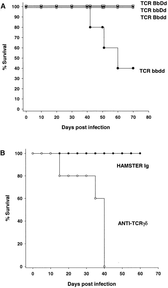
TCR-γ/δ cells regulate HSV-1 infection. (A) TCR null mice were infected with HSV-1 in the hind footpad and were monitored for survival. TCR-β+/−/δ+/− mice, open triangles; TCR-β+/−/δ−/− mice, closed triangles; TCR-β−/−/δ+/− mice, open circles; and TCR-β−/−/ δ−/− mice, closed circles. All groups of mice contained at least three animals, and this plot is representative of two separate experiments. (B) TCR-α−/− mice were infected with HSV-1 in the cornea and were monitored for survival. Control hamster Ig-treated TCR-α−/− mice, closed circles; anti– TCR-γ/δ mAb–treated TCR-α−/− mice, open circles. Both groups of mice contained five animals and this plot is representative of three separate experiments.