
Figure 3.
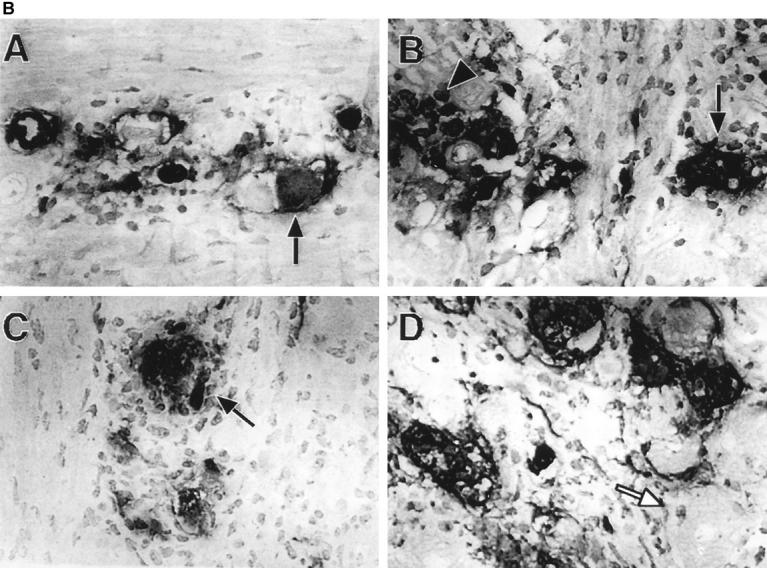

The dynamics of HSV-1 replication is different in TCR-α+/− and TCR-α−/− mice. (A) Quantitation of immunohistochemical staining of HSV-1 viral antigens in the trigeminal ganglia at day 6 after infection. Two mice per group and three representative sections of each ganglia (six sections per group) were prepared for immunohistochemical staining. An average of 1,200 neurons were counted for each group. Data are recorded as the percent of neurons that exhibited specific HSV-1 staining. The differences between the day 6 after infection control and anti–TCR-γ/δ mAb–treated TCR-α+/− mice, or between the control and anti–TCR-γ/δ mAb–treated TCR-α−/− mice, were statistically significant (P <0.05). (B) Photomicrographs depicting immunohistochemical staining of HSV-1 antigens in the trigeminal ganglion. Trigeminal ganglia were obtained 6 d after corneal infection from TCRα+/− mice (A and B) and TCR-α−/− mice (C and D). Mice received control (A and C) or anti–TCR-γ/δ mAb (B and D) treatments. Infected neurons, black arrows; uninfected neurons, white arrows; a cluster of infected inflammatory cells, black arrowhead in B. Original magnification: 100×.