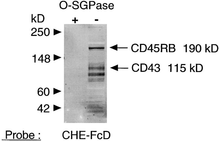
Figure 12.
CD43 and CD45R (exonB) are among the 9-Oacetylated membrane mucins in splenic cells. The left panel shows a Western blot of splenic lymphocyte membrane proteins (15 μg each lane) probed with CHE-FcD (at 4°C) with (lane 1) or without (lane 2) prior treatment of the proteins with OSGPase. The right panel shows a Western blot of total membrane proteins, proteins precipitated by CHE-FcD, and CHE-Fc controls, each probed with antiCD43 and anti-CD45RB monoclonal antibodies as indicated. The expected positions of CD45RB (∼190 kD) and CD43 (∼115 kD) are indicated in each panel. The relatively bright band just above 115 kD in the right lanes of the right panel is nonspecific, since it was also seen on probing with the alkaline phosphatase conjugated secondary reagent alone.


