
Figure 4.
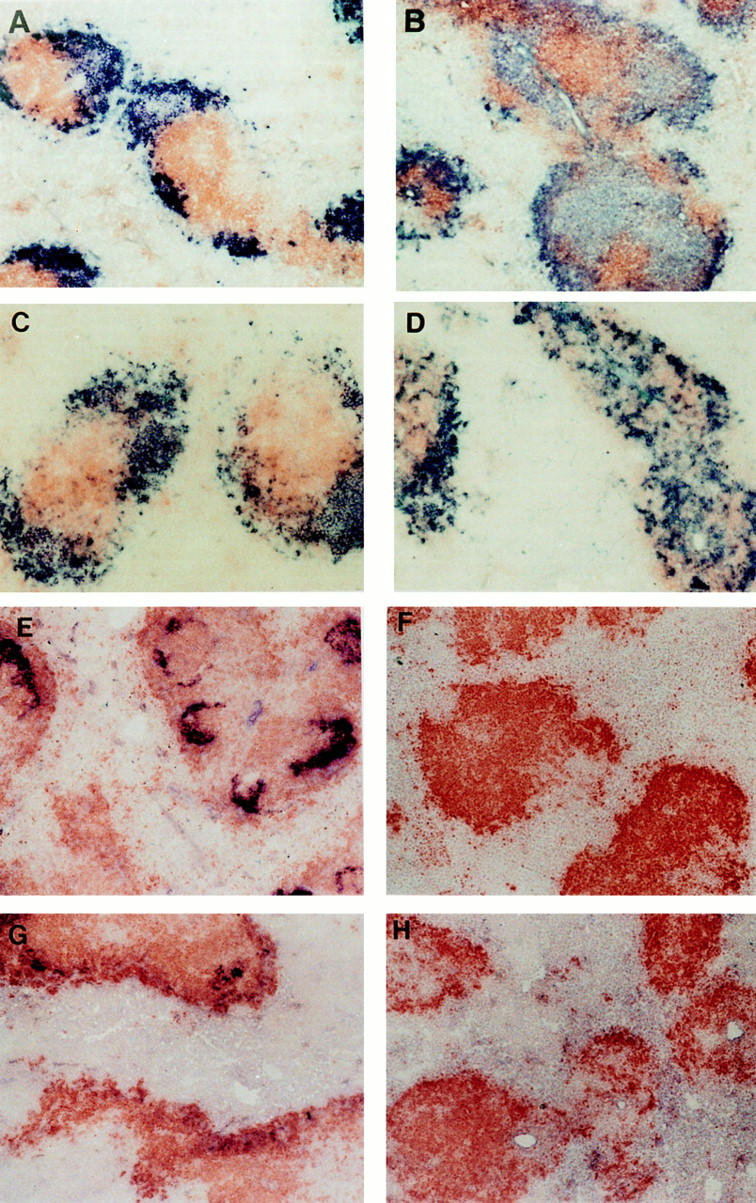
Structure of spleen follicles in irradiated mice reconstituted with wild-type or LTα−/− splenocytes. After serum was collected from mice shown in Fig. 3, the spleens were harvested and frozen sections were stained with anti-B220 (brown) and anti-Thy1.2 (blue) to visualize the B cell and T cell zones (A–D). Distinct B cell and T cell zones were present in wild-type mice that received splenocytes from either normal (A) or LTα−/− mice (C), whereas there was disturbed segregation of B cells and T cells in LTα−/− mice that received splenocytes from either normal (B) or LTα−/− mice (D). FDC clusters were observed by staining with the anti-CR1 monoclonal antibody 8C12 (blue) (E–H). FDC clusters were retained in the spleen follicles of wild-type mice that received splenocytes from either wildtype (E) or LTα−/− mice (G), whereas FDC clusters were absent in the spleens of LTα−/− mice that received splenocytes from either wild-type (F) or LTα−/− mice (H).