
Figure 4.
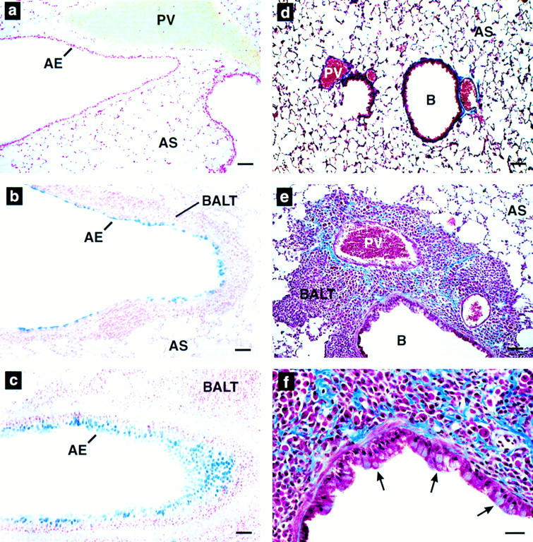
Goblet cell hyperplasia, epithial hypertrophy, and collagen deposition are induced by airway expression of IL-5. (a–c) Alcian blue (pH 2.5) staining of wild-type (a) and NJ.1726 lung sections showing a large bronchus (b) and a smaller more distal bronchiole (c). Intensely blue staining areas are glycoprotein (mucin)-containing goblet cells. (d–e) Masson's trichrome staining of paraffin sections derived from wild-type (d) and NJ.1726 (e and f) lungs. The darkly blue staining extracellular material is collagen. The arrows in f indicate hypertrophy in the bronchial epithelium. AE, airway epithelium; B, bronchiole; PV, pulmonary blood vessel; AS, alveolar space; BALT, bronchus-associated lymphoid tissue. Bars: (a–e) 50 μm; (f) 25 μm..