
Figure 2.
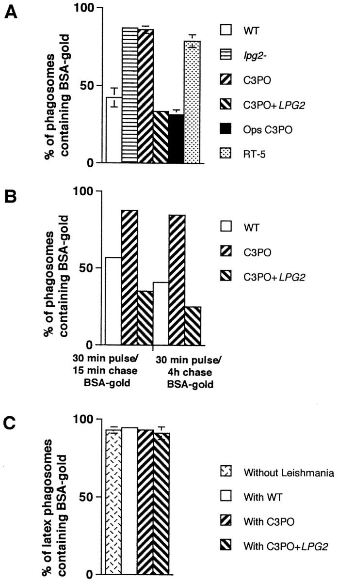
Quantitative analysis of the fusion between phagosomes containing L. donovani parasites and endocytic organelles. (A) Parasites and BSA–gold were internalized as described in Fig. 1. Macrophages were prepared for electron microscopy and quantitative analysis of phagosome– endosome fusion performed on thin sections. The presence of a single gold particle in a parasitophorous vacuole was scored as a fusion event. These results represent the average of three to six experiments. Ops C3PO, LPG-opsonized C3PO. (B) Macrophages were fed a 5- and 16-nm BSA–gold particles mixture for 30 min, followed by either a 15-min or a 4-h incubation period to fill endosomes and lysosomes, respectively. Labeled macrophages were then infected with promastigotes for 60 min and further incubated for 60 min. Fusion was assessed as above. These results represent the average of two experiments. (C) Fusion properties of latex bead–containing phagosomes present in cells coinfected with various parasites (see Fig. 4). Latex bead phagosomes were analyzed only in cells showing the presence of a parasite on the section profile. These results represent the average of three to four experiments.