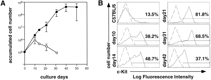
Figure 2.
Growth kinetics and c-Kit expression of BM cells cultured on OP9 stroma cells with mSFO2 containing EGF and SCF. (A) Unfractionated BM cells (105) from C57BL/6 mice were plated on OP9 stroma cells in mSFO2 containing 30 ng/ml EGF (open circle) or 30 ng/ml EGF plus 100 ng/ml SCF (filled circle) in 60-mm culture dishes. On the indicated days, the cultured cells were harvested, counted, analyzed for c-Kit expression, and replated on the fresh OP9 layer. Each point represents the mean of triplicate cultures ± SD. (B) Expression of c-Kit of the cultured cells harvested on the indicated days. Fresh BM (C57BL/6) and cultured BM cells were stained with biotinylated anti–c-Kit (ACK4) antibody revealed by streptavidin-PE. Each staining profile (solid line) was superimposed onto that of the negative control (dashed line) stained with a biotinylated antibody against an irrelevant antigen. Percentage indicates the population of c-Kit–positive cells.

