Figure 3.
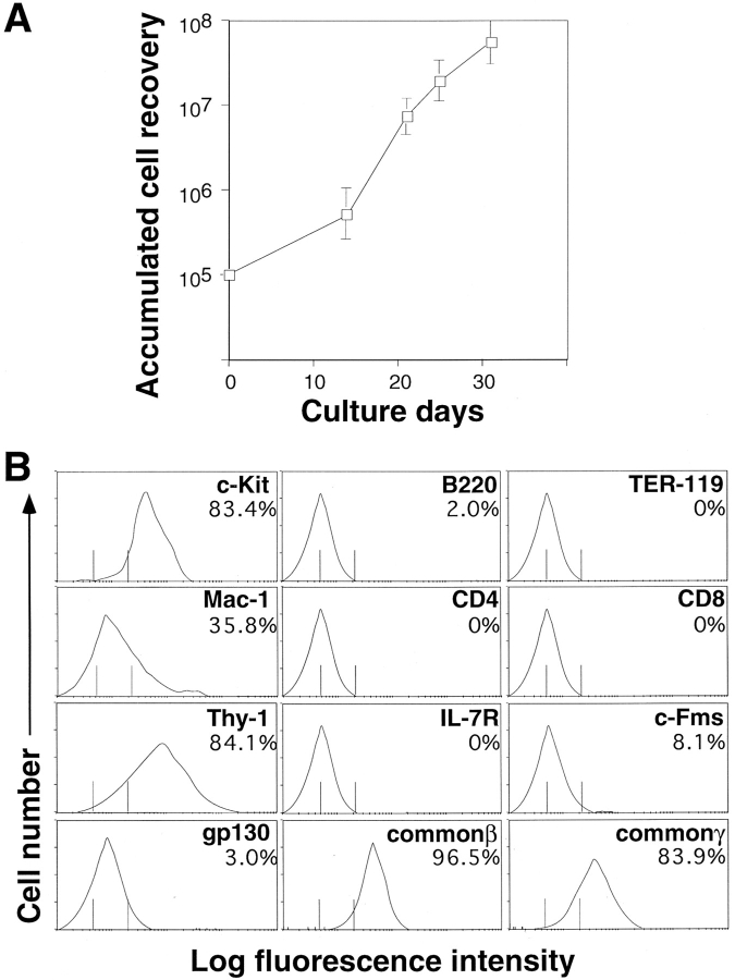
Growth kinetics of BM cells cultured on the OP9/erbB2 stromal layer and surface phenotype of recovered cells. (A) Unfractionated BM cells (105) derived from C57BL/6 were plated on OP9/erbB2 stroma cells in mSFO2 supplemented with 100 ng/ml SCF. On the indicated days, the cultured cells were harvested, counted, and replated onto the fresh OP9/erbB2 stromal layer. (B) Cells harvested on the 21st d of culture were analyzed by flow cytometry after staining with FITCconjugated Mac-1, anti-B220, TER119, CD4, CD8, biotinylated anti–c-Kit (ACK4),Thy-1, anti–IL-7 receptor (A7R), anti–c-Fms (AFS98), antigp130 (GP130), anti–common β (AIC2B), and anti–common γ (TUGm3). Biotinylated antibodies were detected with streptavidin-PE. The vertical lines indicate the mean (left) and the maximal (right) fluorescence intensity of cells stained with an isotype-matched control mAb.

