
Figure 1.
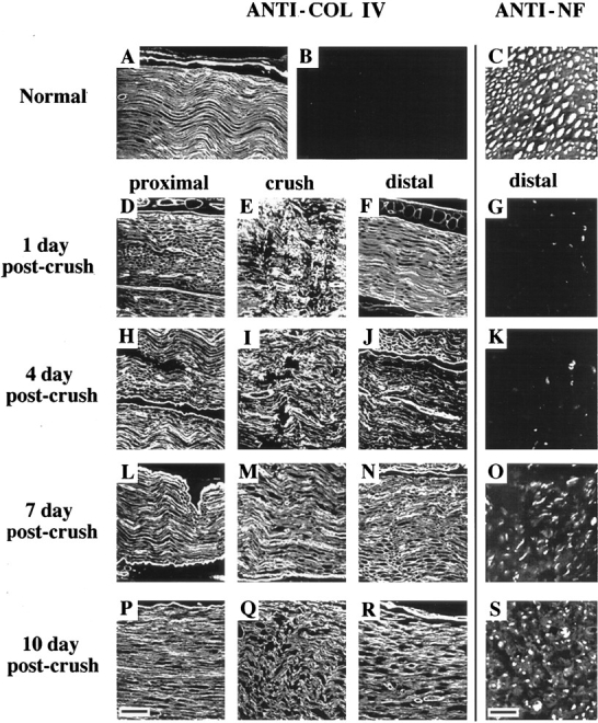
BM integrity and axonal regeneration after sciatic nerve injury. For visualization of BM, longitudinal paraffin sections of uninjured contralateral nerve (A) and injured nerve (proximal, crush, and distal) at 1, 4, 7, and 10 days post-crush were stained with anti-COL IV antibody (D–F, H–J, L–N, and P–R, respectively). (B) A preimmune IgG control. Bar (P), 45 μm. For visualization of axons, transverse paraffin sections of uninjured contralateral nerve (C) and injured nerve approximately 5 mm distal to the crush site at 1, 4, 7, and 10 d after crush (G, K, O, and S, respectively), were stained with anti-neurofilament (ANTI-NF) antibody and visualized by immunofluorescence. Bar (S), 10 μm.