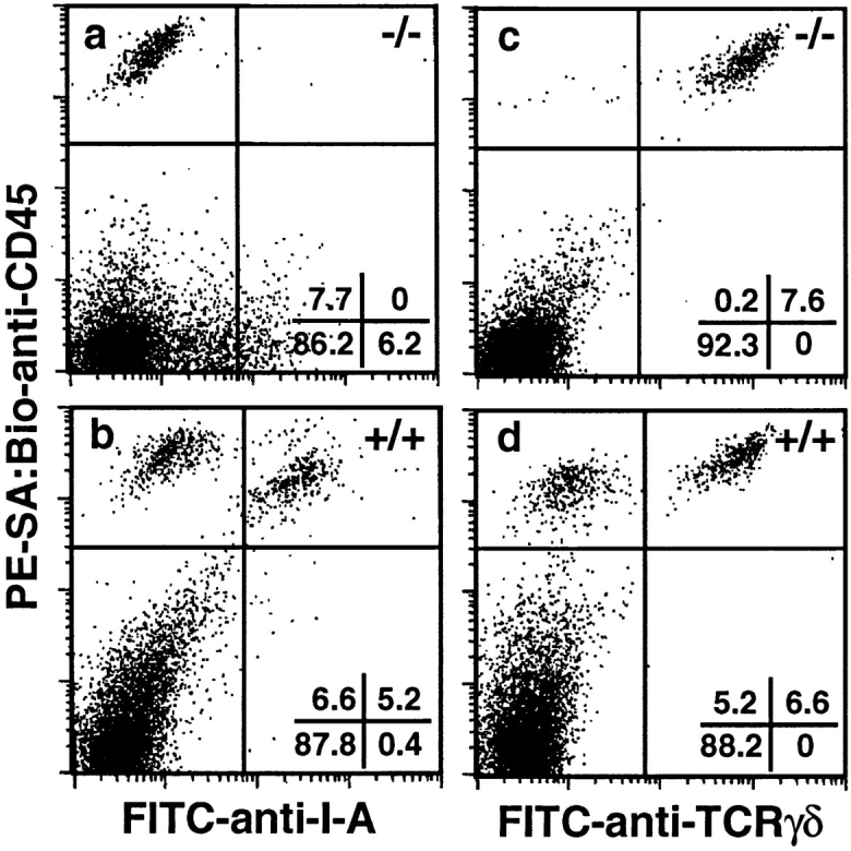
Figure 1.

TGF-β1 −/− mice lack epidermal LC. Epidermal cell suspensions were prepared from the trunk skin of 17-d TGF-β1 −/− and littermate control mice, stained for CD45 and I-A antigens (or CD45 and TCR-γδ), and analyzed using multicolor flow cytometry. Nonviable cells were excluded during data acquisition. Markers were adjusted such that cells stained with isotype control reagents were contained within the left lower quadrant of each panel.
