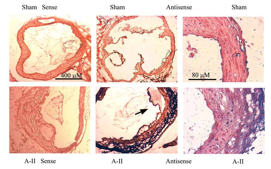
Figure 3.

In Situ hybridization of IL-6 mRNA in aortic cross sections. Aortic cross sections were hybridized with sense (control) and anti-sense IL-6 cRNA probes. Upper panels, Aortae from sham-infused mice probed with sense (far left) and anti-sense IL-6 probes. Two different magnifications are shown (40X and 200X). Lower panels, aortae from A-II-infused mice were hybridized as in the upper panels. Predominant IL-6 staining is in the adventitial layer (purple) with additional diffuse staining in the media and endothelium. Arrow: Endothelial IL-6 staining in region of lesion development.
