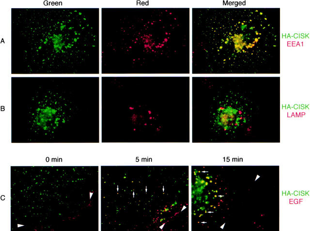
Figure 1.
CISK is localized to endosomal compartments. COS-7 cells transiently transfected with HA-epitope–tagged CISK were examined for the localization of (A) CISK (green) and endogenous EEA1 (red), and (B) CISK (green) and endogenous LAMP (red). Merged images are shown on the right. (C) Texas red–conjugated EGF was added to COS-7 cells expressing HA-CISK. The subcellular localization of CISK (green) and the migration of EGF (red; arrowheads) was examined at 0, 5, and 15 min after EGF addition at 37°C. The migration of EGF with CISK-containing vesicles is indicated by arrows.

