
Figure 4.
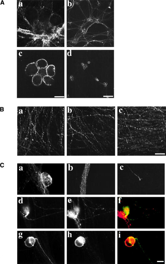
Analysis of the neuronal secretory system during PRV infection. (A) Cell surface gE during wild-type or Us9-null infections. Neurons were infected such that every neuron was infected for 9 h with the wild-type (a and b) or Us9-null virus (c and d). Antibodies that recognize gE were placed directly in the culture medium for 5 min, and then unbound antibodies were removed and the infected neurons were fixed. (B) The steady-state localization of the synaptic vesicle marker SV2. Neurons were mock infected (a) or infected with the wild-type virus (b) or the Us9-null virus (c) such that every neuron was infected for 16 h, and then the neurons were fixed and permeabilized. An antibody that recognizes SV2 was then used in indirect immunofluorescence experiments. (C) The subcellular localization of a cellular protein (NgCAM) expressed during PRV infection. A replication- defective virus expressing the cellular protein NgCAM was used to infect cultured neurons. After 1 h, the medium was removed, and cultures were mock infected (a–c) or infected with the wild-type (d–f) or Us9-null virus (g–i). a–c show NgCAM localization in the absence of PRV infection. NgCAM appears on the surface of the cell body (a), in vesicles within the axon (b; note that this is an image of a large fasiculation, and therefore many axons are present within the bundle), and on the surface of the axon (c). NgCAM labeled the surface of neurons infected with wild-type virus (d) and Us9-null virus (g). The expression of gE during the wild-type (e) and Us9-null (h) infections demonstrates that they are infected with PRV. Merged images of the wild-type infection (f) and Us9-null infection (i) are shown with NgCAM in green and gE in red. Bars: (A, a and b, B, and C) 25 μm; (A, c and d) 150 μm.