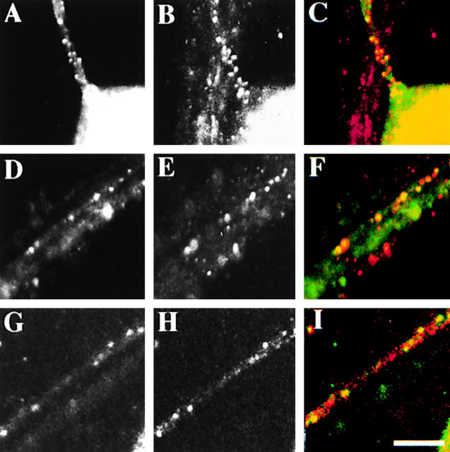
Figure 6.
Colocalization of Us9 with other viral membrane proteins within the axon. Neurons were infected with the wild-type virus such that every neuron was infected for 6 h, and then antibodies to Us9 (A, D, and G) and gB (B), gC (E), or gE (H) were added. The merged images are shown in C, F, and I with Us9 in green and the corresponding membrane protein in red. Bar, 10 μm.

