
Figure 8.
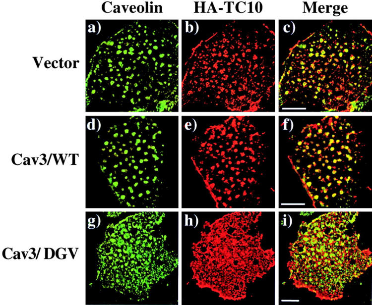
Expression of a dominant- interfering caveolin 3 mutant disrupts the plasma membrane subdomain compartmentalization of TC10. Differentiated 3T3L1 adipocytes were coelectroporated with 50 μg of HA-TC10 plus 200 μg of the empty vector (a–c), wild-type caveolin 3 (Cav3/WT; d–f), or the dominant-interfering caveolin 3 mutant (Cav3/DGV; g–i). 36 h later, the cells were fixed, plasma membrane sheets were prepared and subjected to confocal fluorescent microscopy with a polyclonal caveolin 1 antibody (a, d, and g), and a monoclonal HA antibody (b, e, and h). The merged images are shown in panels c, f, and i. These are representative field of cells from two independent determinations. Bar, 10 μM.