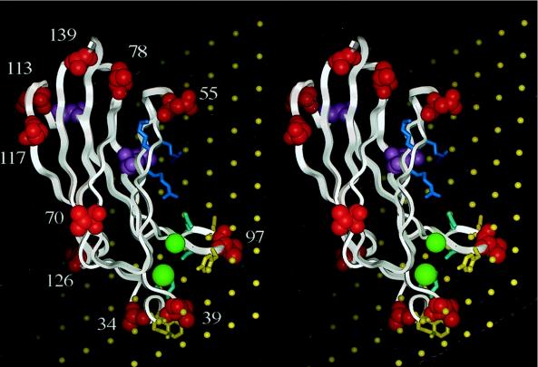
Figure 2.
Stereoview of EPR-based docked C2cPLA2 on DOPM membranes by using the data in Fig. 1. The position of the protein with respect to the membrane (plane of electrostatic potential −77 mV, shown as an array of yellow balls 6 Å apart) is as shown in Fig. 2. The protein backbone is shown as a white ribbon, and native residues that were replaced with spin labels are shown as van der Waals surfaces in red except for residues 88 and 110, which are shown in pink. The two calcium ions are shown as green spheres. CBR1 contains residues 34 and 39 and also F35 and M38 (shown as yellow sticks) and D40 (cyan stick). CBR3 contains V97 and also Y96 and M98 (yellow sticks) and N95 and D99 (cyan sticks). Basic residues R57, K58, and R59 are shown as blue-gray sticks.

