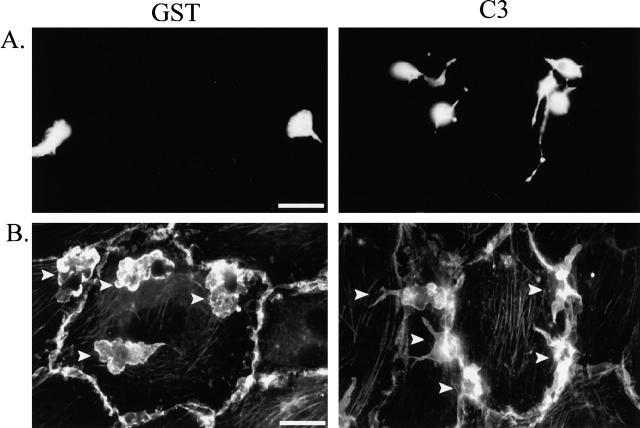
Figure 4.
Morphology of monocytes loaded with C3 when plated on endothelial cells. (A) Either GST- or C3-loaded monocytes were fluorescently labeled with CMFDA and cultured with IL-1–activated endothelial cells for 45 min before fixation. (B) Either GST- or C3-loaded monocytes were cultured with IL-1–activated endothelial cells for 45 min before fixation and staining for F-actin. Arrows indicate the location of monocytes in the cocultures. Bars, 20 μm.

