
Figure 6.
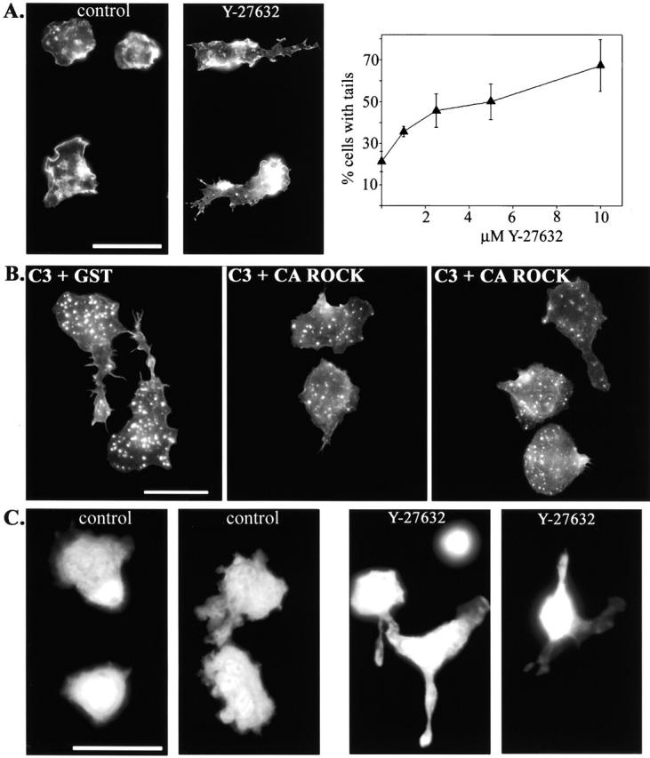
p160ROCK is both necessary and sufficient for RhoA-mediated tail retraction. (A) Monocytes were plated onto coverslips in the presence of 10% autologous serum with or without the p160ROCK inhibitor, Y-27632 (10 μM). The graph represents data obtained from monocytes plated onto coverslips with serum containing media and a 1.0–10 μM range of Y-27632 for 45 min. Cells were then fixed, stained for F-actin, and the percentage of cells with tails was scored. The data plotted represent the average from three separate experiments. After 45 min incubation, cells were fixed and stained for F-actin to reveal cell morphology. (B) Monocytes were electroporated with C3 + CA ROCK and compared with those electroporated with C3 + GST. Cells were plated on coverslips with serum-containing media for 45 min before fixation. Cells were stained for F-actin for morphological assessment. (C) Fluorescently labeled monocytes were pretreated with 10 μM Y-27632 for 15 min and then washed and added to activated endothelial monolayers in the presence of 1 μM Y-27632 for 20 min before fixation. Images reveal the morphology of only the monocytes in the coculture. Cells on the left of each pair of images were on top of the endothelial monolayer, as judged by the focal plane. Cells on the right were underneath the monolayer (controls), or caught between neighboring endothelial cells (Y-27632). Bars, 20 μm.