
Figure 1.
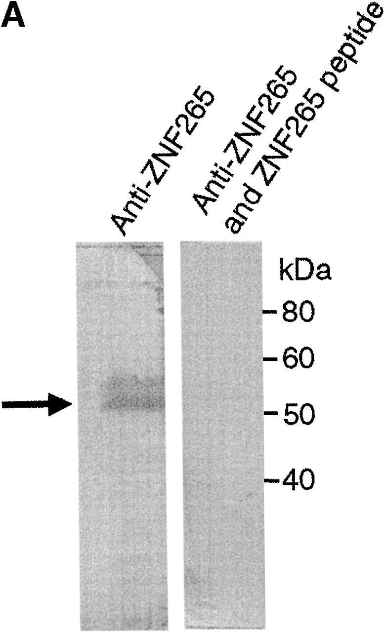

Subcellular colocalization of endogenous ZNF265 with endogenous nuclear factors. (A) Immunoblotting assay demonstrates specific recognition of ZNF265 by the polyclonal ZNF265 antibody (the arrow shows a 55-kD band), which was competed by ZNF265 oligopeptide antigen (2.5 μg/ml) in three replicate experiments. (B) Subcellular localization of various protein factors. Fixed Calu-6 cells were exposed to: (1st column) monoclonal antibodies against splicing factors U1-70K, Sm antigen, SC35, SMN, or transcriptosomal factors p300 and YY1, in respective rows, before incubation with Alexa 594 anti–mouse IgG (red); (2nd column) staining with anti-ZNF265 and detection with Alexa 488–conjugated anti–rabbit IgG (green); (3rd column) DAPI staining of nucleus (blue); (4th column) digital overlay of Z-series projections shown in columns 1 and 2 to demonstrate colocalization (yellow); (5th column) scattergrams of the overlayed projection shown in column 4. Each row represents the same field (width × height = 60 × 60 μm), acquired using three-channel confocal microscopy.