
Figure 5. MMP-9 rescues reduced TIMP-2−/− myotube size without altering β1 integrin expression.
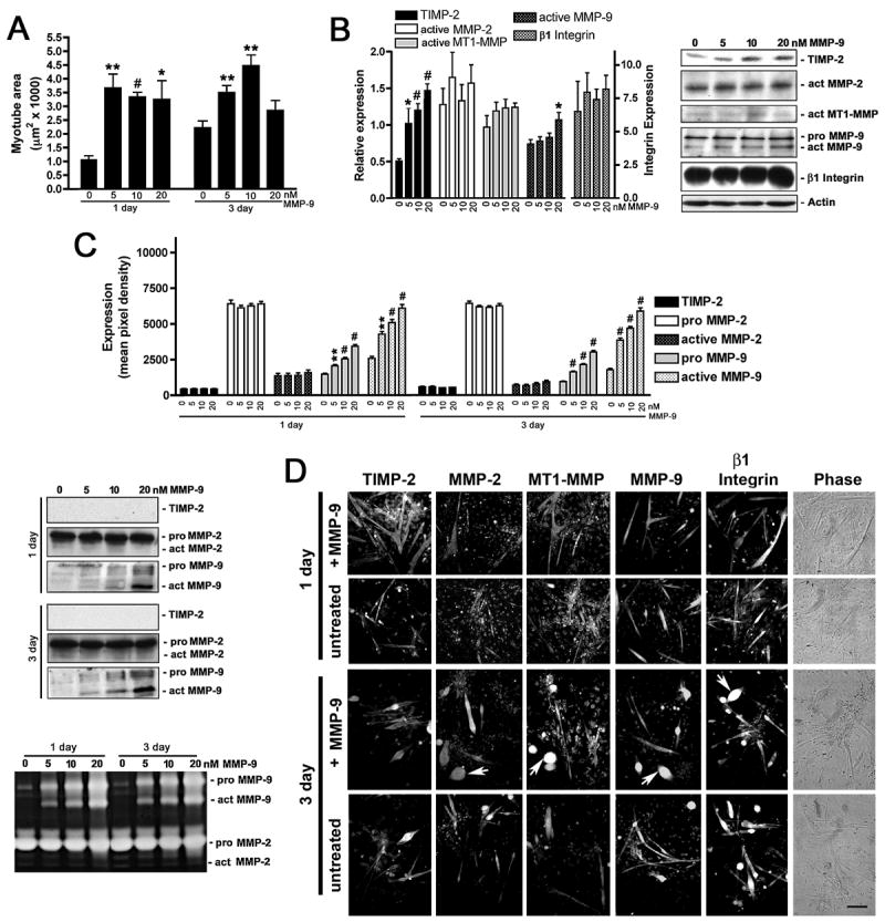
A) TIMP-2−/− myotube area was measured on live cells after 1 and 3 days in differentiation media in the absence or presence of recombinant active mouse MMP-9 (5, 10, 20 nM). MMP-9 rescues TIMP-2−/− myotube size at a lower concentration (5 nM) than MMP-2 (10 nM) at both 1 and 3 days. B) Western blot analysis (25 μg whole cell lysates) shows that MMP-9 treatment increases cellular TIMP-2 expression, but has no effect on β1 integrin expression. C) Western blot and zymographic analysis (25 μl conditioned medium) detects increased active MMP-9 in the medium of MMP-9 treated cells. However, no change in TIMP-2 secretion is detected. D) Immunocytochemistry of Triton permeabilized untreated and MMP-9 treated (10 nM) cells. Note that, like MMP-2 treatment, the number of myoballs (arrows) is increased in MMP-9 treated cultures at 3 days. *p < 0.05, **p ≤ 0.01, #p ≤ 0.001. Scale bar = 100 μm.