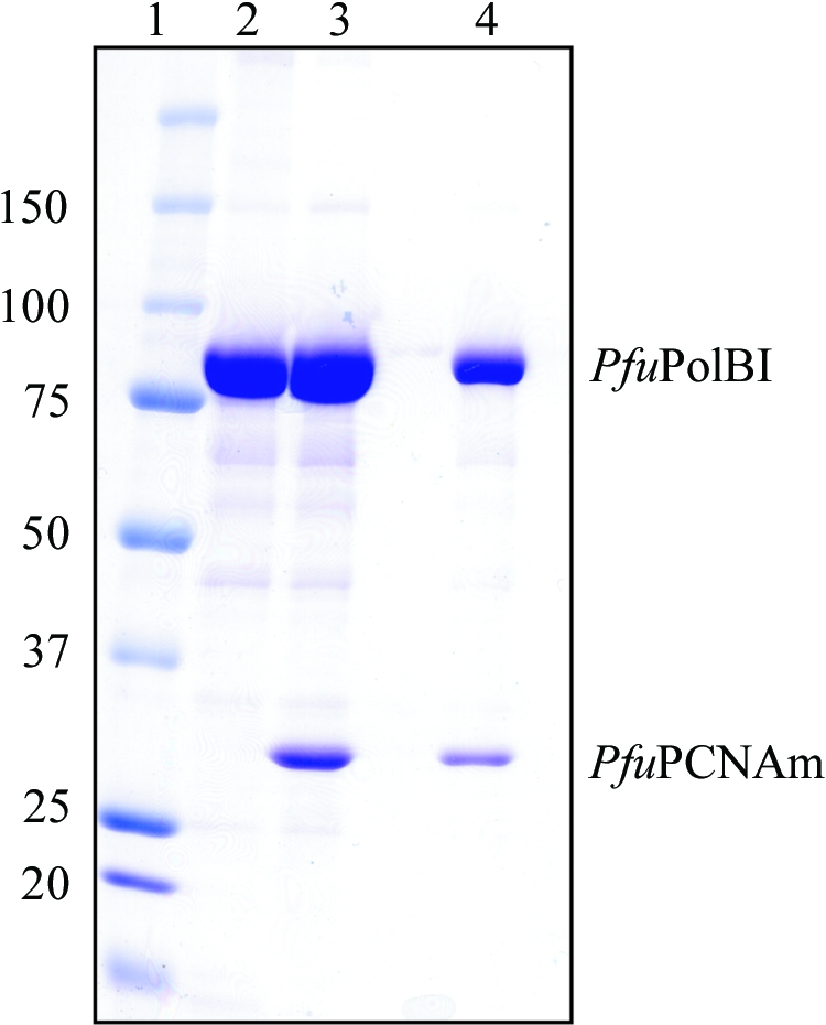
Figure 3.

5 µl aliquots of the peaks from the gel-filtration analysis and a dissolved sample of the obtained crystals were analyzed by SDS–PAGE. The gel was stained with Coomassie blue dye. Lane 1, molecular-weight markers (sizes are shown in kDa on the left); lane 2, PfuPolBI; lane 3, PfuPolBI + PfuPCNAm, gel filtration; lane 4, PfuPolBI + PfuPCNAm, crystal. The crystals were washed in the precipitant solution and then dissolved in buffer A.
