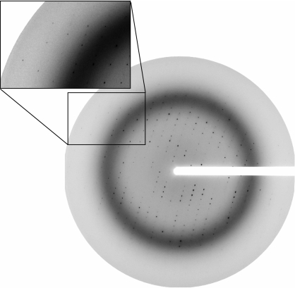
Figure 4.
Diffraction pattern of a ZEBRA175–236/mut–DNA crystal. The crystal was exposed at 100 K after soaking in 25% PEG 400 for cryoprotection. The rotation angle used was 1°, the crystal-to-detector distance was 150 mm and the wavelength was 0.9755 Å. The inset shows a detail of the diffraction pattern with spots visible near the edge of the detector (2.5 Å).

