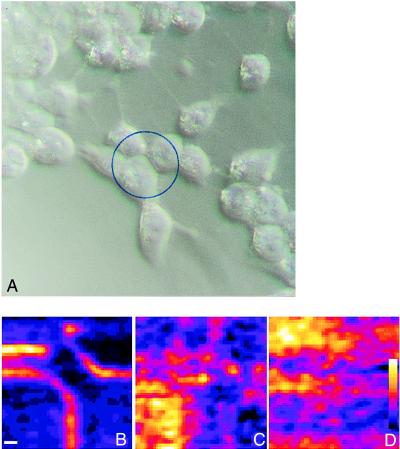
Figure 4.
(A) A conventional optical micrograph of a group of cells that are stained with JPW 1259 and are labeled with the 1-nm gold-conjugated antibody. A portion of the field of view that is indicated by a circle in A was imaged by using second harmonic generation in (B, C, and D). In B, the presence of the nanoparticles on the membranes enhances the second harmonic signal, which traces the membrane outline. Images C and D were obtained during and after an acidic wash was applied, and this caused a disordering of the 1-nm gold-conjugated antibodies. The scale bar in B is 2 microns.

