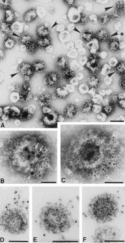
Figure 7.
Localization of GAIP on CCVs after immunogold labeling. (A) CCV fraction negatively stained with 2% uranyl acetate showing enrichment of CCVs with cage-like clathrin coats (black arrowheads). (B and C) Negative staining of CCVs showing labeling for GAIP (5 nm gold) and clathrin (10 nm gold, white arrows). (D–F) A CCV fraction was filtered onto Millipore filters, CCVs were immunolabeled for GAIP with 5 nm gold, anti-rabbit conjugate followed by embedding in Epox and routine electron microscopy. (Bar = 0.1 μm.)

