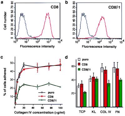
Figure 2.
Effect of CD8β1 on keratinocyte adhesiveness. (a and b) Cell-surface CD8 levels were evaluated by flow cytometry in puro- (black), CD8- (red in a), and CD8β1- (red in b) expressing keratinocytes. Profiles show total population. (c) Cells (104 per well) were plated for 3 hr in the presence of 25 μM cycloheximide. (d) Equal numbers of cells (103 per 35 mm dish) expressing puro, CD8, or CD8β1 were plated overnight on tissue culture plastic (TCP), type IV collagen (COL IV, 50 μg/ml), fibronectin (FN, 25 μg/ml) or keratinocyte extracellular matrix enriched for laminin 5 (KL) in the presence of J2–3T3 feeder cells, and the number of adherent keratinocytes was determined. (Error bars in c and d = standard deviation of the mean of triplicate samples within one experiment.)

