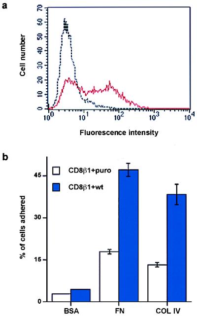
Figure 5.
Introduction of wild-type chicken β1 integrin subunit (wt) into keratinocytes expressing CD8β1. (a) Flow cytometry of basal keratinocytes with JG22 to chicken β1 integrin. Black line: cells expressing CD8β1 and puro. Red line: cells expressing CD8β1 and chicken β1 integrin (CD8β1 + wt). (b) Doubly infected cells (104 ) were plated for 3 hr with 25 μM cycloheximide on heat-denatured BSA (0.5 mg/ml), fibronectin (FN, 25 μg/ml), or type IV collagen (COL IV, 50 μg/ml). (Error bars = standard deviation of the mean of triplicate samples in one experiment.)

