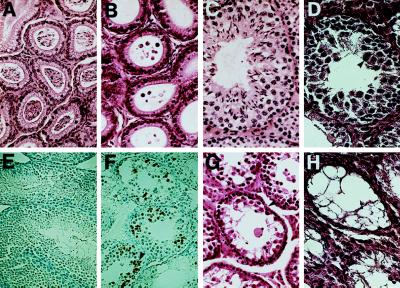
Figure 4.
Histology and immunohistochemical staining of the epididymis of adult sterile and fertile chimeric mice with a Ptn dominant negative mutation. (A and B) Hematoxylin and eosin-stained sections of epididymis of the fertile and sterile animals. The epididymes of the fertile male mice are packed with spermatozoa (A), whereas those of the sterile male mice lacked detectable spermatozoa (B). (C and D) Hematoxylin and eosin-stained section of the fertile and sterile chimeric mouse testes. Normal spermatogenesis was seen in the seminiferous tubules of the 2- to 4-month-old fertile chimeric mice (C), and degeneration of seminiferous epithelium was observed in 2- to 4-month-old sterile chimeric animals generated from Ptn dominant-negative mutation heterozygous E14 or RW4 clones (D). The multinucleated giant cells, as noted by arrowheads, and an expanded layer of cells, indicated by a double-headed arrow, have a nuclear morphology characteristic of premeiotic cells. (E and F) Testis sections of sterile and fertile chimeric mice were stained with the Apop Tag Plus Peroxidase Kit to detect apoptotic cells in situ. The assay shows clusters of apoptotic germ cells and a marked increase in apoptosis in the testes of the sterile mice (F) as compared with the extent of cell death known to occur in normal spermatogenesis (E). The labeled cells are denoted by an arrow. (G and H) In older mutant males, progressive degeneration of the germinal epithelium occurred, ending with the formation of striking vacuolization of tubules that were observed in testis from the sterile males at 6 months (G) and 8 months (H). Sertoli cells and Leydig cells appear normal and are denoted by arrows.

