Figure 2.
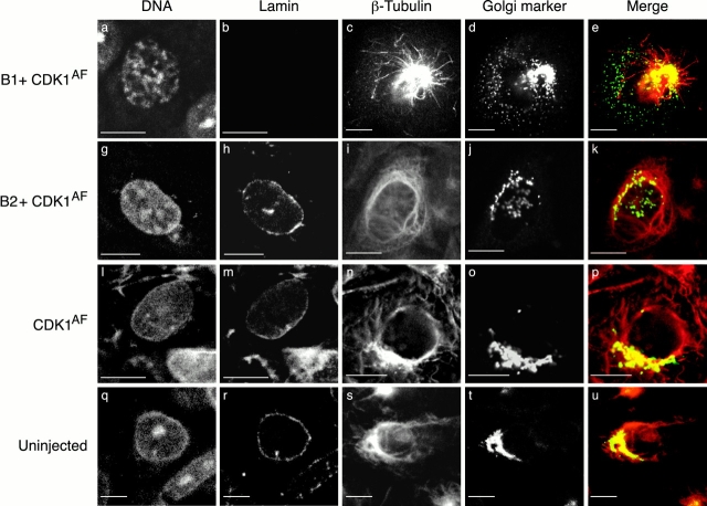
Different effects of cyclin B1– and B2–CDKs on subcellular architecture. Serum-starved CHO cells were unperturbed (bottom row) or microinjected with expression vectors coding for a Golgi marker NAGT–GFP (d, j, and o; green in e, k, and p) and CDK1AF, alone (third row), with cyclin B1 (top row), or with cyclin B2 (second row). After 6 h, the cells were fixed and stained with TOTO-3 to visualize the DNA (a, g, l, and q) and with an antilamin antibody (b, h, m, and r) or an anti–β-tubulin antibody (c, i, n, and s; red in e, k, p, and u). Antimannosidase II was used to stain the Golgi in uninjected cells (t; green in u). Note that cyclin B1 + CDK1AF caused the nuclear lamina to disassemble, and the solubilized lamin protein was washed out of the cell during fixation. The Golgi apparatus fragmented in both cyclin B1–CDK1AF and cyclin B2–CDK1AF injected cells, but only cyclin B1 + CDK1AF caused the microtubules to become much shorter and the centrosome to nucleate more microtubules. Cells are representative of more than 120 cells analyzed in more than three separate experiments. Bars, 10 μm.

