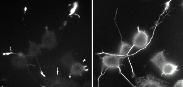
Figure 2.
Immunofluorescent staining of differentiating CAD cells shows the expression and localization of endogenous JIP-1 (left) and tubulin (right) proteins. In cells that have not yet begun to extend neurites (arrow at left), JIP-1 expression and localization are not apparent. But, as soon as this neuron-like cell line has established neurites, JIP-1 is localized to their tips via kinesin-I. The cell denoted by an arrowhead at left is just beginning to produce neurites, whereas the two cells near the center of the field have longer neurites and bright JIP-1 staining at the distal ends. (Image courtesy of K.J. Verhey)

