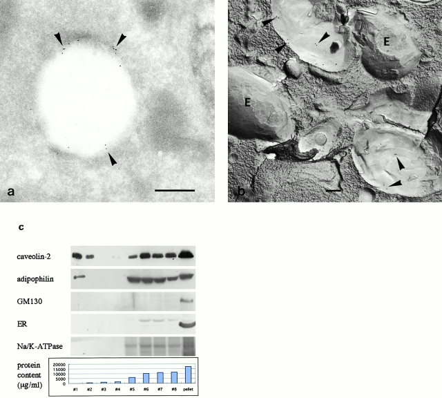
Figure 2.
(a and b) Immunoelectron microscopy of HepG2 expressing caveolin-2β (clone A-8). Bar, 500 nm. (a) Ultrathin cryosection. Gold particles labeling caveolin-2 are observed in the rim of LD in small clusters (arrowheads); the content of LD appears vacant. (b) Freeze-fracture replica. Gold particles for caveolin-2 are observed in clusters (arrowheads) on the P face of LD, which show an onion-like morphology. The E face of LD (E) is devoid of labeling. (c) Western blotting of subcellular fractions obtained from HepG2 clone A-8. The graph shows the protein content of the fractions. The top two fractions (#1 and #2) contained little protein, but showed positive signals for caveolin-2 and adipophilin. GM130, 66-kD protein, and Na+/K+-ATPase (Golgi, ER, and plasma membrane markers, respectively) are found only in the bottom fractions.

