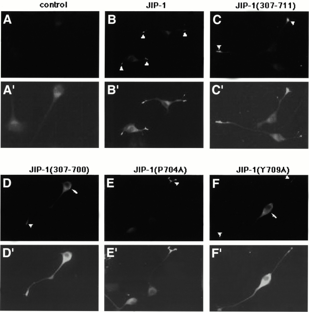
Figure 4.
The COOH-terminal residues of JIP-1 are required for proper subcellular localization. NIE 115 cells were transiently transfected with the parental plasmid (control) or with plasmids encoding the indicated JIP-1 variants, differentiated, and the expressed proteins were detected by indirect immunofluorescence microscopy using an anti-Myc mAb. Nonspecific background staining is visible in the control cells and is enhanced in A′–F′ to aid in visualization of the cells. Myc–JIP-1 variants were scored as positive for correct cellular localization (JIP-1, JIP-1 [307–711], and JIP-1 [P704A]) if fluorescence was more pronounced at the neurite tips (arrowheads), whereas transfected proteins were considered negative for localization (JIP-1 [307–700] and JIP-1 [Y709A]) if fluorescence was observed to be more prominent in the cell body (arrows).

